Our Arterial Ultrasounds
THE EXPERTS IN ARTERIAL ULTRASOUND
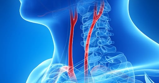
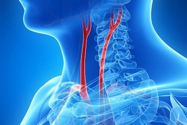
Carotid Doppler
An ultrasound can detect plaque in the arteries that supply blood to the brain. Narrowing of the arteries caused by plaque can lead to stroke a transient ischaemic attack (TIA) or mini stroke. Carotid artery disease often develops slowly and may not cause symptoms until a significant blockage or clot formation occurs.
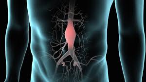
Abdominal Aortic Aneurysm
The aorta and its branches take the blood from the heart down to the abdomen and legs. We can assess for dilatation of the arteries called an aneurysm which often have no symptoms and plaque build up causing narrowing of the artery.

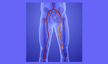
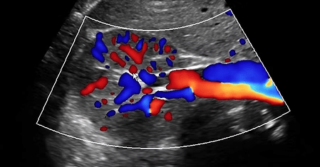
Peripheral Arterial Study
A peripheral arterial ultrasound analyses the flow through the arteries from the abdominal aorta- the main artery off the heart to the arteries at the foot. A blocked artery in the abdomen can stop the flow getting to the calf causing syptoms in the buttock, thigh or calf.
Leg Arteries
Ultrasound is used to assess the arteries of the leg for the presence of atherosclerosis - disease and narrowing of the arteries. Significant disease can cause pain with activity as the demand for blood supply to the leg muscles is increased and unable to be met.


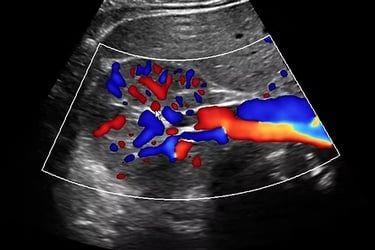
Renal Doppler
A renal Doppler ultrasound is performed to assess the blood flow in the renal arteries, which supply blood to the kidneys. It helps identify any blockages or narrowing of these arteries, which can lead to conditions like renal artery stenosis or high blood pressure.
Mesenteric Arteries
Mesenteric arteries supply blood flow to the intestines. Narrowing of these arteries can occur at any age with loss of the ability to properly digest food, potentially causing pain, nausea, vomiting, diarrhoea and weight loss.


Temporal Arteries
The temporal arteries are vessels located on the sides of the head supplying blood to the scalp, jaw muscles, and optic nerve in the eye. Inflammation of these arteries, known as temporal arteritis or giant cell arteritis, can lead to various symptoms such as headaches, scalp tenderness and potentially serious complications like vision loss.
Arm Arteries
Arterial disease is more complex in the arm with many anatomical variants. Mechanical compression in the thoracic outlet region, vasospasm of the digital arteries, trauma-related thrombi, arteritis, and emboli from the heart or arm aneurysms are pathologies to be considered.


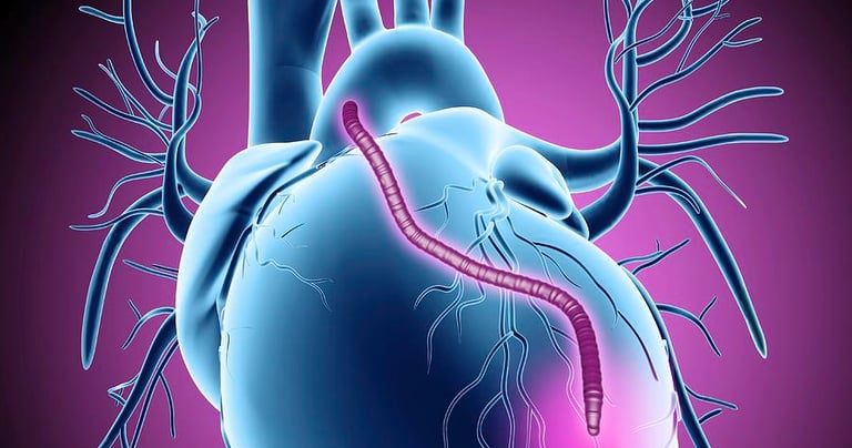
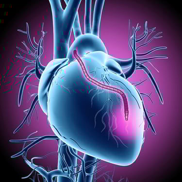
Radial Artery Mapping for CABG
The radial artery is a valuable conduit for a coronary artery bypass graft with excellent long-term patency rates. The ultrasound assessment identifies collateral circulation, which is important to ensure adequate blood flow to the hand after the radial artery is harvested.
Popliteal Entrapment Syndrome
Popliteal entrapment syndrome is a rare condition where the popliteal artery, which supplies blood to the lower leg and foot, is compressed by structures behind the knee, usually muscles. This compression restricts blood flow, causing pain and cramping, especially during exercise.


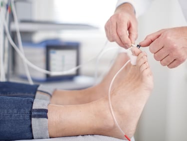
Photoplethysmography
Photoplethysmography (PPG) pressures, are a method of measuring blood pressure in the toes or fingers using a device called a photoplethysmograph. This non-invasive technique helps assess blood flow and peripheral arterial disease, particularly in cases where ankle-brachial index (ABI) testing may be unreliable.


Ankle Brachial Index / + Exercise
The Ankle Brachial Index is a test that compares the blood pressure at the ankles to the arm to assess for peripheral arterial disease. Early detection of artery disease through testing can help individuals receive timely treatment and reduce the risk of complications.